Abstract
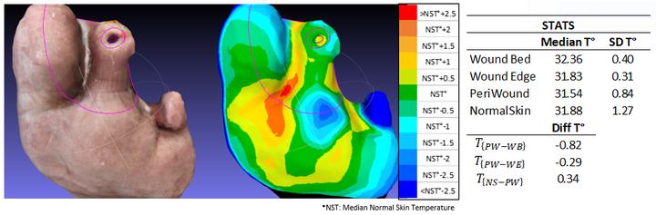
Color, shape (size and volume), and temperature are important clinical features for chronic wound monitoringthat could impact diagnosis and treatment. Noninvasive 3D measurement are better and more accurate thanthose in 2D, but expensive equipment and complexity of the setup prevent their use at hospitals. Therefore,the use of affordable and lightweight devices with straightforward protocol to acquire images for evaluations is fundamental to provide a functional and useful evaluation of the wound. In this work, an automated methodology to generate color and thermal 3D models is presented by using portable devices - a commercial mobile device with a connected portable thermal camera. The 3D model of the wound surface is estimated from a series of color images using structure-from-motion (SfM) while thermal information is overlaid to the ulcer’s relief formultimodal wound evaluation. The proposed methodology contributes with a proof of concept for multimodal wound monitoring in the hospital environment with a simple hand-held shooting protocol. The system was used efficiently with 5 patients on wounds of various sizes and types.
Evelyn Gutierrez
Statistician | Advanced Analytics | R&D | 3D computer vision | Data Scientist
Consultant interested in automation and data-driven decision making